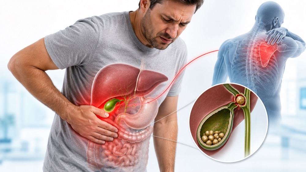
Gallbladder pain has a distinctive signature that sets it apart from other digestive complaints. It strikes predictably after fatty meals, settles in a specific location under your right ribs, and often radiates to your right shoulder blade in a pattern so characteristic that doctors can often diagnose it from the description alone.
But here’s what most articles won’t tell you: Not all gallbladder pain is the same. The discomfort from a temporary stone blockage (biliary colic) feels distinctly different from an infected, inflamed gallbladder (acute cholecystitis)—and knowing which you’re experiencing could mean the difference between scheduling a routine surgery and facing a life-threatening emergency.
This comprehensive guide breaks down exactly what gallbladder pain feels like, why it happens, how to distinguish it from mimics, and the critical warning signs that demand immediate medical attention.
Understanding Your Gallbladder: Why It Causes Pain
Your gallbladder is a small, pear-shaped organ tucked beneath your liver on the right side of your abdomen. Despite its modest size (about 3-4 inches long), it plays a crucial role in digestion.
What the Gallbladder Does
Function: The gallbladder stores and concentrates bile—a digestive fluid produced by your liver that helps break down fats. When you eat, especially fatty foods, your gallbladder contracts (squeezes) to release bile through the cystic duct into the common bile duct, then into the small intestine.
The pain mechanism: Gallbladder pain occurs when this normal flow is blocked. Most commonly, gallstones (hardened deposits of bile components) lodge in the cystic duct, preventing bile from escaping. As the gallbladder continues to contract against this blockage, pressure builds dramatically inside the organ—causing the intense, characteristic pain of biliary colic.
Why Gallbladder Pain Is Often Misdiagnosed
Gallbladder symptoms are notoriously easy to confuse with other conditions:
- Heartburn/GERD: Both cause upper abdominal discomfort after eating
- Peptic ulcers: Both cause burning epigastric pain
- Heart attack: Gallbladder pain can radiate to chest and arm
- Pneumonia: Right lower lobe pneumonia can irritate the diaphragm, causing referred RUQ pain
- Kidney stones: Right-sided stones cause flank pain that can mimic gallbladder radiation
- Appendicitis: Early appendicitis can present with vague upper abdominal pain before localizing to the right lower quadrant
- Functional dyspepsia: Indigestion without structural cause mimics mild gallbladder symptoms
The key differentiator: Gallbladder pain has a distinctive timing pattern (30 minutes to 3 hours after fatty meals), location (right upper quadrant or epigastrium), and radiation (to right shoulder blade or right shoulder tip) that, when present together, are highly specific for gallbladder disease.
Types of Gallbladder Pain: From Mild to Medical Emergency
Gallbladder disease exists on a spectrum. Understanding where your symptoms fall on this spectrum helps determine urgency of care.
1. Biliary Colic (Gallbladder Attack)
What it is: Temporary blockage of the cystic duct by a gallstone. The gallbladder contracts against the obstruction, causing intense pain. The stone typically falls back into the gallbladder or passes through, relieving the obstruction.
Classic presentation:
| Feature | Typical Pattern |
|---|---|
| Onset | Sudden, often 1-3 hours after a fatty meal |
| Location | Right upper quadrant (RUQ) or epigastrium (upper middle abdomen) |
| Radiation | To right shoulder blade (scapula) or right shoulder tip |
| Character | Steady, boring, pressure-like (NOT colicky/cramping despite the name) |
| Intensity | Moderate to severe, often causes patients to pace or seek comfortable position |
| Duration | 30 minutes to 6 hours (typically 1-4 hours) |
| Resolution | Gradual, as stone dislodges |
| Associated symptoms | Nausea (60-70%), vomiting (30-40%), belching, bloating |
| Between attacks | Asymptomatic or mild indigestion |
Key features that distinguish biliary colic:
-
Postprandial timing: Pain reliably follows meals, especially high-fat foods (fried foods, pizza, ice cream, cheese, fatty meats). Many patients report waking up at night with pain after eating a large dinner.
-
Steady, not cramping: Despite being called “colic,” the pain is typically constant and steady rather than wave-like cramping.
-
Position doesn’t help: Unlike kidney stones (where patients can’t find a comfortable position) or musculoskeletal pain (worse with movement), gallbladder pain patients often lie very still.
-
No fever: Biliary colic does NOT cause fever. If fever is present, think acute cholecystitis.
-
Normal between attacks: Patients feel completely well between episodes, though some report fat intolerance or bloating.
2. Acute Cholecystitis (Infected/Inflamed Gallbladder)
What it is: Prolonged blockage of the cystic duct (usually >6 hours) leads to gallbladder inflammation, bacterial overgrowth, and infection. This is a surgical emergency requiring hospitalization.
How it differs from biliary colic:
| Feature | Biliary Colic | Acute Cholecystitis |
|---|---|---|
| Pain duration | <6 hours, self-limited | >6 hours, persistent and worsening |
| Fever | Absent | Present (typically 100.4-102°F / 38-39°C) |
| White blood cell count | Normal | Elevated (>10,000-12,000/mm³) |
| Physical exam | Mild RUQ tenderness | Marked tenderness, guarding, positive Murphy’s sign |
| Systemic symptoms | Nausea/vomiting only | Malaise, anorexia, tachycardia |
| Resolution | Spontaneous | Requires antibiotics and often urgent surgery |
| Complications | None if resolved | Gangrene, perforation, abscess, sepsis |
Murphy’s Sign: A classic physical exam finding for acute cholecystitis. The doctor places fingers under the right rib cage and asks the patient to take a deep breath. If the patient abruptly stops breathing in due to pain (inspiratory arrest), it’s a positive Murphy’s sign—highly specific for acute cholecystitis.
Red flag symptoms of acute cholecystitis:
- Pain lasting more than 6 hours without relief
- Fever or chills
- Persistent vomiting (unable to keep liquids down)
- Marked tenderness to touch in RUQ
- Rapid heart rate (>100 bpm)
- Feeling generally ill or weak
3. Choledocholithiasis (Stone in Common Bile Duct)
What it is: A gallstone has escaped the gallbladder and lodged in the common bile duct, blocking bile flow from the liver to the intestine.
Distinctive features:
- Jaundice: Yellowing of skin and whites of eyes (scleral icterus)
- Dark urine: Tea-colored or cola-colored urine (bilirubin excreted in urine)
- Pale/clay-colored stools: Lack of bile pigment in intestines
- Elevated liver enzymes: Particularly alkaline phosphatase and direct bilirubin
- Pain: Similar to biliary colic but may be more severe and prolonged
Why it’s dangerous: Can lead to ascending cholangitis (bile duct infection)—a life-threatening emergency.
4. Acute Cholangitis (Infected Bile Duct)
What it is: Bacterial infection of the blocked common bile duct. This is a medical emergency with high mortality if not treated promptly.
Charcot’s Triad (present in 50-75% of cases):
- RUQ pain
- Fever (often high, >102°F / 39°C)
- Jaundice
Reynolds’ Pentad (indicates severe, life-threatening cholangitis):
- Charcot’s Triad PLUS:
- Hypotension (low blood pressure, shock)
- Confusion/altered mental status
Mortality rate: Up to 10% even with treatment. Requires emergency ERCP (endoscopic stone removal) and IV antibiotics.
The Classic Gallbladder Pain Pattern: What It Feels Like
Location
Primary location:
- Right upper quadrant (RUQ): The area under your right rib cage, roughly where your right hand would rest if you placed it on your abdomen with your thumb under the ribs
- Epigastrium: Upper middle abdomen, just below the breastbone (more common early in the attack)
Anatomical explanation: The gallbladder sits beneath the liver in the RUQ. Pain originates from distension of the gallbladder wall and inflammation of the surrounding peritoneum.
Why it sometimes starts in the middle: Early gallbladder pain is often referred to the epigastrium because the gallbladder and stomach share similar nerve pathways (visceral afferent fibers via the celiac plexus). As inflammation progresses and involves the parietal peritoneum, pain localizes more specifically to the RUQ.
Radiation Pattern
Classic radiation:
- Right scapula (shoulder blade): Most common and characteristic
- Right shoulder tip: Less common but highly specific
- Right back: Between or below the shoulder blades
- Retrosternal: Can radiate to chest, mimicking heart attack
Why radiation occurs: The gallbladder shares nerve pathways (phrenic nerve, C3-C5 spinal segments) with the shoulder region. Pain signals from the irritated gallbladder are “misinterpreted” by the brain as coming from the shoulder—a phenomenon called referred pain.
Clinical significance: Radiation to the right scapula is present in approximately 50-60% of biliary colic cases and is considered one of the most specific features of gallbladder disease.
Quality and Intensity
Patient descriptions:
- “Someone squeezing my ribs with a vise”
- “A heavy weight sitting under my right ribs”
- “A constant, deep ache that won’t let up”
- “Pressure like a basketball inflated under my ribs”
- “Boring or drilling sensation”
What it’s NOT:
- Sharp, stabbing (more typical of peritonitis or kidney stones)
- Cramping or wave-like (more typical of intestinal colic)
- Burning that moves upward (more typical of GERD)
- Position-dependent (more typical of musculoskeletal pain)
Timing and Triggers
Classic timing:
- Onset: 30 minutes to 3 hours after eating, most commonly 1-2 hours postprandial
- Nighttime attacks: Very common—patients often wake from sleep 2-3 hours after dinner
- Duration: 1-6 hours for biliary colic; persistent and worsening for cholecystitis
Why the delay? It takes time for your body to release cholecystokinin (CCK), the hormone that triggers gallbladder contraction. Fatty foods stimulate the most CCK release, which is why high-fat meals are the most common trigger.
Common trigger foods:
- Fried foods (French fries, fried chicken, donuts)
- High-fat dairy (ice cream, whole milk, cheese, cream)
- Fatty meats (bacon, sausage, ribs, marbled steaks)
- Pizza (combination of fat from cheese and oil)
- Rich desserts (pastries, cakes, chocolate)
- Eggs (particularly yolks, in some patients)
Important note: Some patients report attacks without obvious dietary triggers. This can occur when:
- A stone spontaneously shifts and blocks the duct
- The gallbladder contracts on its own (gallbladders can contract periodically even without food)
- A very large or impacted stone causes chronic symptoms
Associated Symptoms: Beyond the Pain
Gallbladder disease affects more than just pain perception. Understanding associated symptoms helps with recognition.
Gastrointestinal Symptoms
Nausea: Present in 60-70% of biliary colic episodes
- Often builds gradually as pain intensifies
- May persist for several hours after pain resolves
- Can occur independently between attacks in chronic gallbladder disease
Vomiting: Present in 30-40% of episodes
- Typically occurs after pain is well-established
- Does NOT relieve the pain (unlike some GI conditions where vomiting provides relief)
- May contain bile (greenish-yellow fluid)
Bloating and belching: Common but non-specific
- Often attributed to “gas” or “indigestion” by patients
- May be the only symptom in mild or atypical cases
Fat intolerance: Many patients develop aversion to fatty foods
- Learn to avoid trigger foods after repeated attacks
- May report “greasy” or “floating” stools if bile flow is chronically impaired
Systemic Symptoms
Fever:
- NOT present in simple biliary colic
- Suggests acute cholecystitis or cholangitis
- Low-grade (100.4-101°F / 38-38.5°C) in uncomplicated cholecystitis
- High fever (>102°F / 39°C) suggests cholangitis or gangrenous gallbladder
Chills/rigors: Suggest bacteremia (bacteria in bloodstream)—medical emergency
Jaundice:
- Yellow discoloration of skin and sclera (whites of eyes)
- Indicates common bile duct obstruction
- May be accompanied by dark urine and pale stools
- Requires urgent evaluation
Tachycardia: Heart rate >100 bpm suggests systemic inflammation or sepsis
Atypical Presentations: When Gallbladder Disease Doesn’t Follow the Rules
Not all gallbladder disease presents classically. Certain populations and circumstances produce atypical symptoms.
Elderly Patients
Why atypical:
- Diminished pain perception
- Blunted immune response (may not mount fever)
- Multiple comorbidities that confuse the picture
- Communication difficulties (dementia, delirium)
Common presentations:
- Confusion or altered mental status as primary symptom
- Minimal or no pain
- General functional decline (“just not acting right”)
- Sepsis without localizing signs
- Anorexia and weakness
Clinical pearl: Elderly patients have higher rates of gangrenous cholecystitis and perforation precisely because symptoms are subtle and diagnosis is delayed.
Diabetic Patients
Why atypical:
- Diabetic neuropathy can blunt pain perception
- Immune dysfunction increases infection risk
- Higher risk of emphysematous cholecystitis (gas-forming infection)
Concerns:
- May present with DKA (diabetic ketoacidosis) precipitated by infection
- Higher mortality from gallbladder disease
- Require lower threshold for imaging and intervention
Pregnant Women
Why it happens:
- Pregnancy increases cholesterol saturation of bile (more stone formation)
- Progesterone relaxes smooth muscle, impairing gallbladder emptying
- Mechanical pressure from growing uterus
Presentation:
- Similar to non-pregnant patients
- May be confused with normal pregnancy discomforts or preeclampsia
- Right upper quadrant pain in pregnancy should always prompt evaluation for both gallbladder disease and HELLP syndrome/preeclampsia
Management considerations:
- Ultrasound is safe and first-line
- Laparoscopic cholecystectomy is safest in second trimester if needed
- Conservative management preferred if possible
Children and Adolescents
Increasing incidence: Gallstones in children have become more common with rising childhood obesity rates.
Presentation:
- Often less specific than adults
- May present as vague abdominal pain, irritability, or feeding difficulties in young children
- Adolescents typically present similarly to adults
Risk factors in children:
- Obesity
- Hemolytic diseases (sickle cell, hereditary spherocytosis)
- Total parenteral nutrition (TPN)
- Certain medications (ceftriaxone, octreotide)
- Family history
Acalculous Cholecystitis
What it is: Gallbladder inflammation WITHOUT gallstones. Accounts for 5-10% of cholecystitis cases.
Who gets it:
- Critically ill patients (ICU, on mechanical ventilation)
- Major trauma or burn patients
- Prolonged fasting or TPN
- Severe systemic illness (sepsis, vasculitis)
Why it happens: Gallbladder stasis (lack of emptying) combined with ischemia (reduced blood flow) leads to inflammation and infection.
Presentation: Often subtle in critically ill patients—unexplained fever, leukocytosis, or RUQ tenderness in an already-sick patient.
Diagnostic Approach: How Doctors Confirm Gallbladder Disease
Clinical History
Your doctor will ask about:
- Pain characteristics (location, quality, radiation, intensity, duration)
- Timing relative to meals
- Trigger foods
- Associated symptoms (nausea, vomiting, fever, jaundice)
- Previous similar episodes
- Risk factors (age, gender, pregnancy, medications, rapid weight loss)
The “5 F’s” of gallbladder disease (mnemonic for risk factors):
- Female
- Forty (or older)
- Fat (obese)
- Fertile (multiparous—multiple pregnancies)
- Family history
Physical Examination
What your doctor looks for:
- RUQ tenderness: Pain with palpation under right rib cage
- Murphy’s sign: Inspiratory arrest during RUQ palpation on deep breath
- Guarding: Involuntary muscle rigidity over the gallbladder
- Rebound tenderness: Pain when pressure is quickly released (suggests peritoneal inflammation)
- Jaundice: Yellowing of skin or eyes
- Fever: Temperature >100.4°F (38°C)
Laboratory Tests
For biliary colic:
- Often normal
- May show mild leukocytosis (elevated WBC) if prolonged
For acute cholecystitis:
- CBC: Elevated white blood cell count (>10,000-12,000/mm³) with left shift
- CRP: Elevated C-reactive protein (inflammatory marker)
- Liver function tests: May show mild elevation in AST, ALT, alkaline phosphatase, bilirubin
For choledocholithiasis/cholangitis:
- Liver function tests:
- Elevated alkaline phosphatase (cholestatic pattern)
- Elevated direct (conjugated) bilirubin
- Mild-moderate elevation in AST/ALT
- CBC: Markedly elevated WBC
- Coagulation studies: Elevated PT/INR if severe liver dysfunction
- Blood cultures: Often positive in cholangitis
Imaging Studies
Ultrasound (First-line):
- Sensitivity: 85-90% for gallstones, 80-85% for acute cholecystitis
- Findings:
- Gallstones (echogenic foci with posterior acoustic shadowing)
- Gallbladder wall thickening (>3-4 mm)
- Pericholecystic fluid (fluid around gallbladder)
- Sonographic Murphy’s sign (pain when ultrasound probe presses on gallbladder)
- Gallbladder distension
Advantages: No radiation, widely available, inexpensive, can be done at bedside
HIDA Scan (Cholescintigraphy):
- Sensitivity: 95%+ for acute cholecystitis
- How it works: Radioactive tracer injected, taken up by liver, excreted in bile
- Positive for cholecystitis: Tracer enters bile ducts but NOT gallbladder (indicates cystic duct obstruction)
When used: When ultrasound is equivocal or clinical suspicion remains high despite negative ultrasound
CT Scan:
- Sensitivity: 75-85% for gallstones, better for complications
- Findings: Similar to ultrasound plus better visualization of complications (abscess, perforation, gangrene)
When used: When diagnosis is unclear, to evaluate for alternative diagnoses, or to assess complications
MRCP (Magnetic Resonance Cholangiopancreatography):
- Sensitivity: 90-95% for common bile duct stones
- How it works: MRI technique that visualizes bile and pancreatic ducts
- When used: Suspected choledocholithiasis, to plan ERCP
ERCP (Endoscopic Retrograde Cholangiopancreatography):
- Both diagnostic AND therapeutic
- How it works: Endoscope passed into duodenum, catheter inserted into bile duct, contrast injected, X-rays taken
- Can remove stones: Special tools can extract stones from bile duct
When used: Confirmed or highly suspected choledocholithiasis, cholangitis
Treatment Options Based on Diagnosis
Biliary Colic
Immediate management:
- Pain control: NSAIDs (ketorolac, ibuprofen, diclofenac) are first-line; opioids if needed
- Antiemetics: For nausea/vomiting (ondansetron, metoclopramide, promethazine)
- Dietary modification: Avoid fatty foods until definitive treatment
Definitive treatment:
- Laparoscopic cholecystectomy: Gold standard, typically scheduled electively
- Timing: Usually within weeks to months of diagnosis (earlier if frequent attacks)
- Success rate: 90-95% symptom resolution
Why surgery is recommended:
- 70% of patients will have recurrent attacks within 2 years
- Risk of complications (cholecystitis, pancreatitis, cholangitis) is 1-2% per year
- Each attack carries risk of progression to more serious condition
Acute Cholecystitis
Hospital management:
- IV fluids: For hydration and hemodynamic support
- IV antibiotics: Cover gram-negative and anaerobic organisms (ceftriaxone + metronidazole, piperacillin-tazobactam, ampicillin-sulbactam)
- Pain control: IV NSAIDs and/or opioids
- NPO: Nothing by mouth (gallbladder rest)
Surgical management:
- Early laparoscopic cholecystectomy: Within 72 hours of symptom onset (preferred approach)
- Delayed cholecystectomy: After 6-8 weeks if initial conservative management successful
- Open cholecystectomy: If laparoscopic approach fails or not feasible
If patient is too unstable for surgery:
- Percutaneous cholecystostomy: Drainage tube placed through skin into gallbladder (interventional radiology)
- Used as bridge to surgery in critically ill patients
Choledocholithiasis
Management:
- ERCP with sphincterotomy and stone extraction: First-line for common bile duct stones
- Followed by cholecystectomy: To prevent future stones (typically within same hospitalization or within 2 weeks)
If ERCP fails:
- Surgical common bile duct exploration: Laparoscopic or open
- Percutaneous transhepatic cholangiography (PTC): Alternative approach
Acute Cholangitis
Medical emergency requiring:
- IV antibiotics: Broad-spectrum, high-dose
- IV fluids: Aggressive resuscitation
- Biliary decompression: Usually via ERCP within 24-48 hours
- ICU care: If severe (hypotension, altered mental status)
Mortality: Up to 10% even with treatment; significantly higher if untreated
Complications of Untreated Gallbladder Disease
Gangrenous Cholecystitis
What it is: Gallbladder tissue death from prolonged ischemia (lack of blood flow).
Risk factors:
- Delayed presentation (>72 hours of symptoms)
- Elderly patients
- Diabetes
- Cardiovascular disease
Consequences:
- High risk of perforation
- May require open surgery instead of laparoscopic
- Higher mortality rate
Gallbladder Perforation
What it is: Hole develops in gallbladder wall, spilling infected bile into abdomen.
Types:
- Free perforation: Into peritoneal cavity → generalized peritonitis, sepsis
- Contained perforation: Walled off by surrounding organs → pericholecystic abscess
Mortality: 15-30% even with treatment
Treatment: Emergency surgery, drainage of abscess, IV antibiotics
Gallstone Pancreatitis
What it is: Gallstone passes through common bile duct and obstructs pancreatic duct.
Frequency: Accounts for 40-70% of acute pancreatitis cases
Severity: Can range from mild to life-threatening (necrotizing pancreatitis)
Treatment: Supportive care for pancreatitis, ERCP if persistent bile duct obstruction, cholecystectomy during same hospitalization
Gallbladder Cancer
Association: Long-standing gallstones (especially large stones >3cm) increase risk
Risk level: Absolute risk remains low (0.3-3% in patients with gallstones)
Presentation: Often asymptomatic until advanced; may present as “cholecystitis” that doesn’t improve with treatment
Prevention and Risk Reduction
Modifiable Risk Factors
Maintain healthy weight:
- Obesity increases cholesterol secretion into bile
- BUT avoid rapid weight loss (>3 lbs/week), which increases stone formation
- Gradual weight loss (1-2 lbs/week) with balanced diet
Dietary modifications:
- Limit refined carbohydrates and sugar
- Choose healthy fats (olive oil, fish, nuts) over saturated fats
- Increase fiber intake (fruits, vegetables, whole grains)
- Stay well-hydrated
Exercise regularly:
- Physical activity improves gallbladder emptying
- Associated with reduced gallstone risk
Medication review:
- Some medications increase stone risk (estrogen therapy, fibrates, octreotide, ceftriaxone)
- Discuss alternatives with doctor if high risk
Non-Modifiable Risk Factors
Cannot change, but should increase awareness:
- Female gender
- Age >40
- Family history
- Certain ethnicities (Native American, Hispanic)
- Personal history of gallstones
When to Seek Medical Attention
Call Your Doctor If:
- First episode of RUQ or epigastric pain lasting >1 hour
- Recurrent episodes of postprandial pain
- Pain severe enough to limit activities
- Persistent nausea or vomiting after meals
- Known gallstones with new or worsening symptoms
Go to Emergency Department If:
- Pain lasting >6 hours without relief
- Fever >100.4°F (38°C) with abdominal pain
- Yellowing of skin or eyes (jaundice)
- Dark urine and pale stools
- Persistent vomiting (unable to keep liquids down)
- Severe pain with abdominal tenderness to touch
- Confusion, weakness, or fainting
- Rapid heart rate (>100 bpm) with abdominal pain
Call 911 If:
- Signs of septic shock (confusion, very low blood pressure, inability to stay awake)
- Severe abdominal pain with rigid, board-like abdomen
- Unconsciousness or near-unconsciousness
Frequently Asked Questions
Can gallbladder pain go away on its own?
Biliary colic episodes typically resolve within 1-6 hours as the stone dislodges. However, the underlying gallstones remain, and recurrent attacks are very common (70% within 2 years). Acute cholecystitis will NOT resolve on its own and requires medical treatment.
What’s the difference between gallbladder pain and heartburn?
Gallbladder pain is typically more severe, located in RUQ or epigastrium, radiates to the right shoulder blade, follows fatty meals by 1-3 hours, and doesn’t respond to antacids. Heartburn is typically a burning sensation that moves upward from the epigastrium, responds to antacids, and is often worse when lying down.
Can you have gallbladder pain without gallstones?
Yes—this is called biliary dyskinesia or functional gallbladder disorder. The gallbladder doesn’t empty properly despite no stones being present. Diagnosis requires a HIDA scan showing low ejection fraction (<35-40%). Treatment is still cholecystectomy.
Is gallbladder surgery a major surgery?
Laparoscopic cholecystectomy is minimally invasive but still considered major surgery. Most patients go home the same day or after one night. Recovery is typically 1-2 weeks for desk work, 4-6 weeks for heavy lifting. Open cholecystectomy (needed in 5-10% of cases) requires longer recovery (6-8 weeks).
Can you live without a gallbladder?
Yes, completely normally. Your liver still produces bile—it just drips continuously into the intestine rather than being stored and concentrated. Most people have no long-term dietary restrictions after recovery. Some experience temporary diarrhea or fat intolerance that usually resolves within months.
Are there non-surgical treatments for gallstones?
Ursodiol (ursodeoxycholic acid): Can dissolve small cholesterol stones in select patients, but takes months to years, and stones recur in 50% within 5 years. Only works for radiolucent (cholesterol) stones in functioning gallbladder.
Shock wave lithotripsy: Breaks up stones with sound waves, but high recurrence rate and limited availability.
Bottom line: Surgery is the only definitive cure for symptomatic gallstones.
What happens if I delay gallbladder surgery?
You risk recurrent painful attacks and potential complications (cholecystitis, pancreatitis, cholangitis). Each attack carries the risk of progression to emergency, which has higher morbidity than elective surgery. However, if you have infrequent, mild attacks and prefer to avoid surgery, watchful waiting is reasonable after discussing risks with your doctor.
The Bottom Line: Know Your Gallbladder Pain
Gallbladder disease is one of the most common surgical conditions, affecting 10-15% of adults. The hallmark features—right upper quadrant pain occurring 1-3 hours after fatty meals, radiating to the right shoulder blade, lasting 1-6 hours—are distinctive enough that patients and doctors can often recognize it from the history alone.
Key takeaways:
-
Biliary colic (temporary blockage) causes intense but self-limited pain without fever. It’s not an emergency but requires surgical consultation.
-
Acute cholecystitis (infected gallbladder) causes persistent pain (>6 hours) with fever and tenderness. It’s urgent—requiring hospitalization and often emergency surgery.
-
Jaundice + fever + pain suggests bile duct obstruction with possible infection (cholangitis)—a life-threatening emergency requiring immediate intervention.
-
Ultrasound is the first-line diagnostic test. Combined with clinical presentation and basic labs, it can confirm the diagnosis in most cases.
-
Laparoscopic cholecystectomy is the definitive treatment—safe, effective, and usually curative. Most patients recover fully and live normally without a gallbladder.
Don’t ignore the warning signs. Gallbladder disease rarely improves on its own, and delaying treatment can turn a routine surgery into a life-threatening emergency. If your symptoms match the classic pattern described here, see your doctor for proper evaluation.
Last Updated: March 10, 2026
Sources
-
NIDDK. Gallstones. National Institute of Diabetes and Digestive and Kidney Diseases. 2020. https://www.niddk.nih.gov/health-information/digestive-diseases/gallstones
-
Mayo Clinic. Gallstones. Mayo Clinic. 2025. https://www.mayoclinic.org/diseases-conditions/gallstones/symptoms-causes/syc-20354214
-
NHS. Gallstones. National Health Service. 2023. https://www.nhs.uk/conditions/gallstones/
-
Portincasa P, Di Ciaula A, Bonfrate L, et al. Management of common biliary stones: A narrative review. Journal of Clinical Medicine. 2021;10(5):1047. doi:10.3390/jcm10051047
-
Gurusamy KS, Davidson C, Gluud C, Davidson BR. Early versus delayed laparoscopic cholecystectomy for people with acute cholecystitis. Cochrane Database of Systematic Reviews. 2013;(6):CD005440. doi:10.1002/14651858.CD005440.pub3
-
Strasberg SM. Acute calculous cholecystitis. New England Journal of Medicine. 2008;358(26):2804-2811. doi:10.1056/NEJMra0708074
-
American College of Surgeons. Surgical treatment of gallstones. ACS. 2022. https://www.facs.org/for-medical-professionals/education/tools-and-resources-for-educators/sesap-18/surgical-treatment-of-gallstones/
-
Lammert C, Gurusamy K, Ko CW, et al. Gallstones. Nature Reviews Disease Primers. 2016;2:16024. doi:10.1038/nrdp.2016.24
-
Tazuma S, Unno M, Igarashi Y, et al. Evidence-based clinical practice guidelines for cholelithiasis 2016. Journal of Gastroenterology. 2017;52(3):276-300. doi:10.1007/s00535-016-1289-7
-
Shabanzadeh DM, Sørensen LT, Jørgensen T. A prediction rule for gallstone disease. BMC Gastroenterology. 2017;17:76. doi:10.1186/s12876-017-0633-7
-
UpToDate. Acute calculous cholecystitis in adults. 2024. https://www.uptodate.com/contents/acute-calculous-cholecystitis-in-adults
-
Society of American Gastrointestinal and Endoscopic Surgeons (SAGES). Guidelines for the clinical application of laparoscopic biliary tract surgery. 2020. https://www.sages.org/publications/guidelines/