Gallstones: Causes, Symptoms, Diagnosis, and Treatment Options Explained
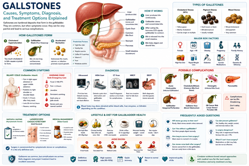
Gallstones are one of the most common digestive disorders in developed countries, affecting 10-15% of adults in the United States—that’s over 25 million Americans. Yet despite their prevalence, many people don’t understand what causes them, when they become dangerous, or what treatment options actually exist.
Most gallstones cause no problems and are discovered incidentally during imaging for other conditions. But when they do cause symptoms, gallstones can be extremely painful and potentially life-threatening if complications develop.
This comprehensive guide explains everything you need to know about gallstones: what they are, why they form, how to recognize the symptoms, how they’re diagnosed, and what treatment options are available—from watchful waiting to surgery.
What Are Gallstones?
Gallstones (cholelithiasis) are hardened deposits that form in the gallbladder, a small pear-shaped organ located beneath your liver on the right side of your abdomen. The gallbladder stores and concentrates bile—a digestive fluid produced by your liver that helps break down fats.
Composition of Gallstones
Gallstones aren’t all the same. They’re classified by their primary composition:
Cholesterol Gallstones (80% in Western countries):
- Color: Yellow-green
- Composition: Primarily undissolved cholesterol
- Size: Range from sand-grain sized to golf balls (average is pea-sized)
- Number: Can be single or multiple (often multiple)
- Risk factors: Female gender, obesity, high-fat diet, rapid weight loss, certain medications
Pigment Gallstones (20% in Western countries):
- Color: Dark brown or black
- Composition: Excess bilirubin (a breakdown product from red blood cells)
- Size: Usually small
- Number: Often multiple
- Risk factors: Cirrhosis, hemolytic anemias (sickle cell, thalassemia), bile duct infections, older age
Mixed Gallstones:
- Contain both cholesterol and pigment components
- Very common—many “cholesterol” stones have pigment centers
- Often form when cholesterol stones become infected
Size and Number Variations
Gallstones vary enormously:
- Size: From 1-2mm (sand-like, called “gallbladder sludge” or microlithiasis) to 8-10cm (large enough to fill the entire gallbladder)
- Number: Single stones or hundreds of small stones
- Shape: Round, faceted (multiple flat surfaces from pressing against each other), or irregular
Clinical significance:
- Small stones (<5mm) are more likely to escape into bile ducts, causing pancreatitis or cholangitis
- Large stones (>3cm) are associated with higher gallbladder cancer risk
- Multiple small stones often cause more symptoms than single large stones
How Gallstones Form: The Underlying Mechanisms
Understanding why gallstones form requires understanding bile composition and gallbladder function.
Bile Composition: A Delicate Balance
Bile contains three key components that must remain in balance:
- Cholesterol (what can form stones)
- Bile acids/bile salts (keep cholesterol dissolved)
- Phospholipids/lecithin (also keep cholesterol dissolved)
Think of bile like a glass of water with dissolved sugar. If you add more sugar than the water can hold, crystals form. Similarly, if bile contains more cholesterol than bile acids and lecithin can keep dissolved, cholesterol crystals form—and these can grow into stones.
Three Mechanisms of Stone Formation
1. Cholesterol Supersaturation (Most Common)
What happens: The liver secretes too much cholesterol into bile relative to bile acids and phospholipids.
Why it occurs:
- Increased hepatic cholesterol secretion (obesity, high-calorie diet, insulin resistance)
- Decreased bile acid production (genetic factors, certain medications)
- Decreased lecithin secretion
Result: Cholesterol precipitates out of solution, forming microscopic crystals that can grow into visible stones.
2. Gallbladder Hypomotility (Sluggish Emptying)
What happens: The gallbladder doesn’t empty regularly or completely, allowing bile to become overly concentrated.
Why it occurs:
- Prolonged fasting (skip meals regularly)
- Very low-calorie diets
- Pregnancy (progesterone relaxes smooth muscle, including gallbladder)
- Rapid weight loss
- Total parenteral nutrition (TPN—IV feeding)
- Spinal cord injury
- Certain medications (octreotide, ceftriaxone)
Result: Stagnant bile becomes concentrated, cholesterol crystals have time to form and aggregate into stones.
3. Accelerated Nucleation
What happens: Certain factors promote faster crystal formation even when cholesterol levels aren’t dramatically elevated.
Contributing factors:
- Mucin hypersecretion (gallbladder lining produces excess glycoproteins that trap cholesterol)
- Presence of calcium salts (form nuclei for crystal growth)
- Bacterial infection (bacteria produce enzymes that deconjugate bilirubin, forming pigment stones)
- Inflammation
Result: Crystals form more rapidly than normal, leading to stone development.
Timeline of Stone Formation
Gallstones don’t form overnight:
- Weeks 1-4: Bile becomes supersaturated with cholesterol
- Weeks 4-12: Microscopic crystals begin to form
- Months 3-6: Crystals aggregate into visible stones (2-5mm)
- Months 6-24: Stones grow larger (5-15mm typical)
- Years 2-10: Stones may continue growing, may become symptomatic
Important: Many people have gallstones for years or decades without ever developing symptoms.
Risk Factors: Who Gets Gallstones?
Gallstone formation is influenced by a complex interplay of genetic, environmental, and lifestyle factors.
The “5 F’s” (Classic Mnemonic)
Medical students learn the “5 F’s” of gallstone risk:
- Female – Women are 2-3 times more likely than men to develop gallstones
- Forty (or older) – Risk increases with age
- Fat (obese) – Excess weight increases cholesterol secretion
- Fertile (multiple pregnancies) – Pregnancy increases stone risk
- Family history – Genetics play a significant role
This mnemonic captures the major risk factors but isn’t comprehensive.
Non-Modifiable Risk Factors
Gender:
- Women: 20% develop gallstones by age 60
- Men: 8% develop gallstones by age 60
- Why: Estrogen increases cholesterol secretion; progesterone reduces gallbladder emptying
Age:
- Rare under age 20
- Incidence doubles every decade after age 40
- By age 75: 25% of women, 10-12% of men have gallstones
Ethnicity:
- Native Americans: Highest prevalence (60-75% in some groups, especially Pima Indians)
- Hispanic/Latino: Higher risk (25-30%)
- Caucasian: Intermediate risk (10-15%)
- African American: Lower risk (5-8%)
- Asian American: Lower overall risk, but higher pigment stone prevalence
Genetics/Family History:
- Having a first-degree relative with gallstones doubles your risk
- Twin studies suggest 25-30% of risk is genetic
- Specific gene variants identified (ABCG8, ABCB4) affect cholesterol transport
Medical Conditions:
- Cirrhosis – Increases pigment stone risk
- Hemolytic anemias – Sickle cell disease, thalassemia, hereditary spherocytosis (increased bilirubin from red blood cell breakdown)
- Crohn’s disease – Especially with ileal resection (bile acid malabsorption)
- Cystic fibrosis – Altered bile composition
- Spinal cord injury – Reduced gallbladder motility
- Organ transplant recipients – Immunosuppressive medications
Modifiable Risk Factors
Obesity:
- BMI >30: 2-3x increased risk vs. normal weight
- Each 1-unit BMI increase = 3-4% increased risk
- Mechanism: Increased hepatic cholesterol secretion
Diet:
- High refined carbohydrates (white bread, sugar, pasta)
- High saturated and trans fats
- Low fiber intake
- Low physical activity
Rapid Weight Loss:
- Losing >3 lbs/week dramatically increases risk
- 10-25% of rapid weight loss patients develop gallstones
- Mechanism: Mobilized tissue cholesterol + reduced gallbladder emptying
- Highest risk: Weeks 2-16 of weight loss
Medications:
- Estrogen therapy (HRT, oral contraceptives) – Increases cholesterol secretion
- Fibrates (cholesterol-lowering) – Increase cholesterol excretion into bile
- Ceftriaxone (antibiotic) – Forms complexes with calcium in bile
- Octreotide – Reduces gallbladder contractility
- Thiazide diuretics – May concentrate bile
Pregnancy:
- Each pregnancy increases risk
- Progesterone reduces gallbladder emptying
- Estrogen increases cholesterol secretion
- Risk persists postpartum (stones formed during pregnancy may remain)
Protective Factors
Some factors REDUCE gallstone risk:
Coffee consumption:
- 2-3 cups daily: 25-40% lower risk
- Both caffeinated and decaf are protective
- Mechanism: Caffeine stimulates gallbladder contraction
Physical activity:
- Regular exercise (150+ min/week): 20-25% lower risk
- Mechanism: Improves insulin sensitivity, weight control
High fiber diet:
- 25-35g fiber daily: 10-17% lower risk
- Mechanism: Binds bile acids, forcing liver to use cholesterol for new bile acid production
Moderate alcohol intake:
- 1-2 drinks daily: 20-30% lower risk (but risks may outweigh benefits)
- Mechanism: May increase HDL, reduce cholesterol saturation
Vitamin C:
- Adequate intake associated with lower risk
- Mechanism: Required for cholesterol → bile acid conversion
Nuts:
- Regular consumption (5+ times/week): 25% lower risk
- Mechanism: Healthy fats, fiber, magnesium
Symptoms: When Gallstones Cause Problems
Asymptomatic Gallstones (Silent Stones)
The majority (60-80%) of gallstones cause NO symptoms.
Key facts:
- Discovered incidentally during ultrasound, CT, or MRI for other reasons
- Most people with asymptomatic stones NEVER develop symptoms
- Annual risk of developing symptoms: 1-2% per year
- 10-year risk of developing symptoms: 10-20%
- 20-year risk: ~20-30%
Management: No treatment needed. “Watchful waiting” is appropriate. Prophylactic cholecystectomy is NOT recommended for asymptomatic stones except in specific high-risk situations (see below).
When prophylactic surgery MAY be considered:
- Gallbladder polyps >1cm
- Porcelain gallbladder (calcified wall—high cancer risk)
- Very large stones (>3cm)
- Sickle cell disease (symptoms hard to distinguish from sickle crises)
- Before bariatric surgery or organ transplant
- Native American ethnicity (very high complication rates)
Symptomatic Gallstones
When gallstones cause symptoms, it’s typically because a stone temporarily or permanently blocks bile flow.
Biliary Colic (Gallbladder Attack)
What it is: Temporary blockage of the cystic duct by a gallstone. The gallbladder contracts against the obstruction, causing pain. The stone typically falls back or passes through, relieving the blockage.
Classic presentation:
| Feature | Typical Pattern |
|---|---|
| Onset | Sudden, 1-3 hours after fatty meal |
| Location | Right upper quadrant or epigastrium |
| Radiation | To right shoulder blade or right shoulder |
| Character | Steady, pressure-like, boring (NOT colicky) |
| Intensity | Moderate to severe |
| Duration | 30 minutes to 6 hours (typically 1-4 hours) |
| Associated symptoms | Nausea (60-70%), vomiting (30-40%) |
| Resolution | Gradual, spontaneous |
Key characteristics:
- Postprandial timing: Reliably follows meals, especially high-fat foods
- Nighttime attacks: Common—patients wake from sleep 2-3 hours after dinner
- Position doesn’t help: Patients often lie very still
- No fever: Biliary colic does NOT cause fever
- Normal between attacks: Patients feel well between episodes
Frequency:
- After first attack: 50% have another within 1 year, 70% within 2 years
- Attacks may become more frequent and severe over time
Complications of Gallstones
When stones cause prolonged blockage or escape into bile ducts, serious complications can develop.
1. Acute Cholecystitis (Infected Gallbladder)
What it is: Prolonged cystic duct obstruction (>6 hours) leads to gallbladder inflammation and bacterial infection.
Frequency: 15-20% of patients with symptomatic gallstones
Symptoms:
- Pain lasting >6 hours (persistent, worsening)
- Fever (100.4-102°F / 38-39°C)
- Marked RUQ tenderness
- Nausea, vomiting
- Malaise, loss of appetite
- Elevated white blood cell count
Diagnosis: Ultrasound (gallbladder wall thickening, pericholecystic fluid, sonographic Murphy’s sign)
Treatment: Hospitalization, IV antibiotics, cholecystectomy (typically within 72 hours)
2. Choledocholithiasis (Stone in Common Bile Duct)
What it is: Gallstone escapes gallbladder and lodges in common bile duct.
Frequency: 10-15% of patients with gallstones
Symptoms:
- Biliary colic-type pain
- Jaundice (yellow skin/eyes)
- Dark urine (tea-colored)
- Pale/clay-colored stools
- Elevated liver enzymes (alkaline phosphatase, direct bilirubin)
Diagnosis: Ultrasound, MRCP, or EUS (endoscopic ultrasound)
Treatment: ERCP to remove stone, followed by cholecystectomy
3. Acute Cholangitis (Infected Bile Duct)
What it is: Bacterial infection of the obstructed common bile duct.
Frequency: Less common but life-threatening
Charcot’s Triad (50-75% of cases):
- RUQ pain
- Fever (often high, >102°F)
- Jaundice
Reynolds’ Pentad (severe, life-threatening):
- Charcot’s Triad PLUS:
- Hypotension (shock)
- Confusion/altered mental status
Mortality: Up to 10% even with treatment; requires emergency ERCP and IV antibiotics
4. Gallstone Pancreatitis
What it is: Gallstone obstructs pancreatic duct (where it joins common bile duct).
Frequency: Accounts for 40-70% of acute pancreatitis cases
Symptoms:
- Severe epigastric pain radiating to back
- Nausea, vomiting
- Elevated amylase and lipase
Treatment: Supportive care, ERCP if persistent obstruction, cholecystectomy during same hospitalization
5. Gallbladder Empyema
What it is: Pus-filled gallbladder from severe infection.
Symptoms: High fever, severe pain, markedly elevated WBC
Treatment: Emergency cholecystectomy or cholecystostomy drainage
6. Gallbladder Gangrene and Perforation
What it is: Prolonged obstruction leads to tissue death and hole in gallbladder wall.
Risk factors: Elderly, diabetes, delayed presentation (>72 hours)
Consequences: Peritonitis, abscess, sepsis
Mortality: 15-30% even with treatment
7. Gallstone Ileus
What it is: Large gallstone erodes through gallbladder into intestine, causing bowel obstruction.
Rare: <1% of gallstone complications
Treatment: Surgery to remove stone
8. Gallbladder Cancer
Association: Long-standing gallstones (especially >3cm) increase risk
Absolute risk: Low (0.3-3% in patients with gallstones)
Presentation: Often asymptomatic until advanced
Treatment: Radical surgery (often not curative by time of diagnosis)
Diagnosis: How Gallstones Are Detected
Clinical History and Physical Exam
History questions:
- Pain characteristics (location, quality, radiation, intensity, duration)
- Timing relative to meals
- Trigger foods
- Associated symptoms (nausea, vomiting, fever, jaundice)
- Previous similar episodes
- Risk factors
Physical exam findings:
- RUQ tenderness: Pain with palpation under right ribs
- Murphy’s sign: Inspiratory arrest during RUQ palpation on deep breath (specific for acute cholecystitis)
- Guarding: Involuntary muscle rigidity
- Jaundice: Yellowing of skin or eyes
- Fever: Temperature >100.4°F (38°C)
Laboratory Tests
For uncomplicated biliary colic:
- Often normal
- May show mild leukocytosis
For acute cholecystitis:
- CBC: Elevated WBC (>10,000-12,000/mm³)
- CRP: Elevated (inflammatory marker)
- Liver enzymes: May show mild elevation
For choledocholithiasis/cholangitis:
- Liver function tests:
- Elevated alkaline phosphatase
- Elevated direct bilirubin
- Mild-moderate AST/ALT elevation
- CBC: Markedly elevated WBC
- Coagulation studies: May be elevated if severe liver dysfunction
- Amylase/lipase: If pancreatitis suspected
- Blood cultures: Often positive in cholangitis
Imaging Studies
Transabdominal Ultrasound (First-Line)
Advantages:
- No radiation
- Widely available
- Inexpensive
- Bedside capability
- High accuracy for gallstones
Findings:
- Gallstones (echogenic foci with posterior acoustic shadowing)
- Gallbladder wall thickening (>3-4mm)
- Pericholecystic fluid
- Sonographic Murphy’s sign
- Gallbladder distension
- Common bile duct dilation
Accuracy:
- Sensitivity: 85-90% for gallstones
- Sensitivity: 80-85% for acute cholecystitis
HIDA Scan (Cholescintigraphy)
When used: Ultrasound equivocal but high clinical suspicion
How it works: Radioactive tracer injected, taken up by liver, excreted in bile
Positive for cholecystitis: Tracer enters bile ducts but NOT gallbladder (cystic duct obstruction)
Accuracy: 95%+ for acute cholecystitis
CT Scan
When used:
- Diagnosis unclear
- Evaluating for complications (abscess, perforation, gangrene)
- Assessing alternative diagnoses
Findings: Similar to ultrasound plus better visualization of complications
Accuracy:
- Sensitivity: 75-85% for gallstones
- Better for complications than simple stones
MRCP (Magnetic Resonance Cholangiopancreatography)
When used: Suspected common bile duct stones
How it works: MRI technique visualizing bile and pancreatic ducts
Accuracy: 90-95% for common bile duct stones
Advantages: Non-invasive, no radiation
EUS (Endoscopic Ultrasound)
When used: Suspected small common bile duct stones not seen on other imaging
How it works: Ultrasound probe on endoscope passed into duodenum
Accuracy: 95%+ for common bile duct stones
ERCP (Endoscopic Retrograde Cholangiopancreatography)
Both diagnostic AND therapeutic:
- Endoscope passed into duodenum
- Catheter inserted into bile duct
- Contrast injected, X-rays taken
- Stones can be removed during same procedure
When used:
- Confirmed or highly suspected choledocholithiasis
- Acute cholangitis (emergency)
- Failed other stone removal techniques
Risks: Pancreatitis (3-5%), bleeding, perforation, infection
Treatment Options
Treatment depends on whether stones are asymptomatic or symptomatic, and whether complications are present.
Asymptomatic Gallstones
Standard approach: Watchful waiting (no treatment)
Rationale:
- 60-80% never develop symptoms
- Annual risk of developing symptoms: only 1-2%
- Risks of surgery outweigh benefits in asymptomatic patients
Exceptions (prophylactic cholecystectomy may be considered):
- Gallbladder polyps >1cm
- Porcelain gallbladder
- Very large stones (>3cm)
- Sickle cell disease
- Before bariatric surgery or organ transplant
- Native American ethnicity
Symptomatic Gallstones (Biliary Colic)
Definitive Treatment: Laparoscopic Cholecystectomy
What it is: Surgical removal of the gallbladder using minimally invasive laparoscopic technique.
Why it’s recommended:
- 70% will have recurrent attacks within 2 years
- Risk of complications: 1-2% per year
- Each attack carries risk of progression to more serious conditions
Procedure details:
- General anesthesia
- 4 small incisions (5-10mm each)
- Camera and instruments inserted
- Gallbladder dissected from liver bed
- Cystic duct and artery clipped and divided
- Gallbladder removed through one incision
- Duration: 1-2 hours typically
Outcomes:
- Success rate: 95%+ symptom resolution
- Most patients go home same day or after one night
- Return to normal activities: 1-2 weeks (desk work), 4-6 weeks (heavy labor)
Complications (uncommon):
- Bile duct injury (0.3-0.5%)
- Bleeding (1-2%)
- Infection (1-3%)
- Bile leak (0.5-1%)
- Conversion to open surgery (5-10%)
- Death (<0.1% in elective cases)
Timing:
- Elective (scheduled): Within weeks to months of diagnosis
- Early (within 72 hours of acute cholecystitis onset): Preferred if patient stable
Medical Management (for patients who cannot or will not have surgery)
Ursodiol (Ursodeoxycholic Acid):
- Mechanism: Reduces cholesterol secretion into bile
- Dosing: 8-10 mg/kg/day in 2-3 divided doses
- Duration: 6-24 months
- Success rate: 60-70% for small stones (<1.5cm)
- Recurrence rate: 50% within 5 years after stopping
- Best candidates: Small cholesterol stones, functioning gallbladder, poor surgical candidates
Extracorporeal Shock Wave Lithotripsy (ESWL):
- Mechanism: Sound waves break up stones
- Success rate: Variable; high recurrence
- Limited availability: Rarely used now
Contact dissolution:
- Mechanism: Solvent (MTBE) injected directly into gallbladder
- Success rate: Variable
- Risks: Significant; rarely used
Dietary management:
- Low-fat diet may reduce attack frequency
- Does NOT eliminate stones or prevent complications
- Appropriate only for patients awaiting surgery or who are poor surgical candidates
Acute Cholecystitis
Hospital Management:
- IV fluids: Hydration and hemodynamic support
- IV antibiotics: Cover gram-negative and anaerobes
- Ceftriaxone + metronidazole
- Piperacillin-tazobactam
- Ampicillin-sulbactam
- Duration: 4-7 days typically
- Pain control: IV NSAIDs (ketorolac) and/or opioids
- NPO: Nothing by mouth (gallbladder rest)
Surgical Management:
Early laparoscopic cholecystectomy (within 72 hours):
- Preferred approach
- Shorter hospital stay
- Lower complication rate
- Definitive treatment
Delayed cholecystectomy (6-8 weeks later):
- If initial conservative management successful
- If patient unstable for immediate surgery
- Higher risk of recurrent attacks while waiting
If Patient Too Unstable for Surgery:
Percutaneous cholecystostomy:
- Drainage tube placed through skin into gallbladder
- Done by interventional radiology
- Used as bridge to surgery in critically ill patients
- Can be removed once patient stabilizes and definitive surgery performed
Choledocholithiasis (Common Bile Duct Stones)
Management:
- ERCP with sphincterotomy and stone extraction:
- First-line for common bile duct stones
- Success rate: 90-95%
- Risks: Pancreatitis (3-5%), bleeding, perforation
- Followed by cholecystectomy:
- To prevent future stones
- Typically within same hospitalization or within 2 weeks
- Rationale: Gallbladder is source of stones
If ERCP Fails:
- Surgical common bile duct exploration (laparoscopic or open)
- Percutaneous transhepatic cholangiography (PTC)
Acute Cholangitis
Medical Emergency Requiring:
- IV antibiotics: Broad-spectrum, high-dose
- Piperacillin-tazobactam
- Cefepime + metronidazole
- Carbapenems (if severe or resistant organisms suspected)
- IV fluids: Aggressive resuscitation
- Biliary decompression: Within 24-48 hours
- ERCP preferred
- PTC if ERCP not available or fails
- Surgery if other methods fail
- ICU care: If severe (hypotension, altered mental status)
Mortality: Up to 10% even with treatment; significantly higher if untreated
Living Without a Gallbladder
What Changes After Cholecystectomy?
Immediate post-operative period (first 2-4 weeks):
- Bile drips continuously into intestine (not stored/concentrated)
- May experience diarrhea (10-20% of patients)
- Fat intolerance with large fatty meals
- Bloating, gas
Long-term (3+ months):
- Most patients adapt completely
- Bile ducts slightly dilate to take over storage function
- 80-90% have no long-term dietary restrictions
- 10-20% have some ongoing fat sensitivity
- <5% have chronic diarrhea requiring medication
Post-Cholecystectomy Diet
Weeks 1-2:
- Very low fat (<20g/day)
- Small, frequent meals (6 per day)
- Bland, easily digestible foods
- Avoid: fried foods, fatty meats, full-fat dairy, gas-producing vegetables
Weeks 3-4:
- Gradually increase fat to 30-40g/day
- Introduce one new food every 2-3 days
- Monitor tolerance
Month 2+:
- Most patients return to normal diet
- Continue emphasizing fiber and healthy fats
- Some remain fat-sensitive long-term
If diarrhea persists:
- Soluble fiber supplementation (psyllium)
- Bile acid binder medication (cholestyramine, colesevelam)
- Limit very high-fat meals
Post-Cholecystectomy Syndrome
What it is: Persistent or recurrent symptoms after gallbladder removal.
Frequency: 10-40% report some symptoms (most mild and self-limited)
Causes:
- Retained common bile duct stones (2-5%)
- Bile duct injury or stricture
- Sphincter of Oddi dysfunction
- Functional bowel disorders (IBS, functional dyspepsia)
- Other GI conditions (GERD, peptic ulcer disease)
- Non-GI causes (musculoskeletal pain, cardiac)
Evaluation:
- Liver function tests
- Ultrasound or MRCP
- EUS if high suspicion of retained stones
- ERCP if intervention needed
Treatment:
- Depends on underlying cause
- ERCP for retained stones or sphincter of Oddi dysfunction
- Medications for functional disorders
- Reassurance for mild, non-specific symptoms
Prevention
While not all gallstone risk factors are modifiable, several lifestyle changes can reduce risk:
Maintain Healthy Weight
Obesity increases risk, but:
- Avoid rapid weight loss (>3 lbs/week)
- Aim for gradual loss (1-2 lbs/week)
- Include moderate fat at meals during weight loss (prevents stasis)
Dietary Modifications
Increase:
- Fiber (25-35g daily): Whole grains, fruits, vegetables, legumes
- Healthy fats: Olive oil, nuts, fish, avocados
- Plant protein: Beans, lentils, tofu
- Calcium: Low-fat dairy, leafy greens
Decrease:
- Refined carbohydrates: White bread, sugar, pasta
- Saturated fats: Red meat, butter, full-fat dairy
- Trans fats: Processed foods, fried foods
Regular Physical Activity
- 150+ minutes moderate aerobic activity weekly
- Plus 2 days strength training
- Associated with 20-25% lower gallstone risk
Coffee Consumption
- 2-3 cups daily associated with 25-40% lower risk
- Both caffeinated and decaf protective
Medication Considerations
- Discuss alternatives if on medications that increase risk (estrogen, fibrates, octreotide)
- Ursodiol prophylaxis during rapid weight loss or post-bariatric surgery
Frequently Asked Questions
Can gallstones go away on their own?
Spontaneous dissolution is extremely rare. Small stones may occasionally pass into the intestine without causing symptoms, but this is unpredictable and potentially dangerous (can cause pancreatitis). Most gallstones persist indefinitely without treatment.
How long does gallbladder surgery take to recover from?
- Hospital stay: Same day or 1 night (laparoscopic)
- Return to desk work: 1-2 weeks
- Return to heavy lifting/manual labor: 4-6 weeks
- Full recovery: 6-8 weeks
Open surgery (needed in 5-10% of cases) requires longer recovery (6-8 weeks for normal activities).
Can you live a normal life without a gallbladder?
Yes, completely. Your liver still produces bile—it just drips continuously into the intestine rather than being stored. Most people have no long-term restrictions. Some experience temporary diarrhea or fat intolerance that usually resolves within months.
What happens if gallstones are left untreated?
Asymptomatic stones: Usually nothing—most never cause problems.
Symptomatic stones: Recurrent painful attacks are likely (70% within 2 years). Risk of complications (cholecystitis, pancreatitis, cholangitis) is 1-2% per year. Complications can be life-threatening if not treated promptly.
Are there non-surgical treatments for gallstones?
Ursodiol (prescription medication) can dissolve small cholesterol stones in select patients, but takes 6-24 months, and stones recur in 50% within 5 years after stopping. Shock wave lithotripsy exists but has high recurrence rates. Surgery is the only definitive cure.
Can diet prevent gallstones?
Yes, to some extent. High-fiber diets, moderate healthy fat intake, limited refined carbohydrates, and maintaining healthy weight all reduce risk. However, genetics and non-modifiable factors (gender, age, ethnicity) play significant roles.
Is gallbladder surgery dangerous?
Laparoscopic cholecystectomy is one of the most common surgeries performed worldwide and is very safe in experienced hands. Serious complications occur in <5% of elective cases. Death is extremely rare (<0.1%). Risks are higher for emergency surgery and in patients with significant comorbidities.
What are the first signs of gallbladder problems?
Typical early symptoms include:
- Pain in right upper abdomen or upper middle abdomen
- Pain occurring 1-3 hours after fatty meals
- Pain radiating to right shoulder blade
- Nausea after eating
- Bloating, belching
If you have these symptoms, see your doctor for evaluation.
The Bottom Line
Gallstones are extremely common, affecting 10-15% of adults. Most cause no problems and require no treatment. When they do cause symptoms, however, gallstones can be very painful and potentially dangerous.
Key takeaways:
- Asymptomatic gallstones generally don’t require treatment—watchful waiting is appropriate.
- Symptomatic gallstones typically require laparoscopic cholecystectomy, which is safe, effective, and curative in 95%+ of cases.
- Complications (acute cholecystitis, cholangitis, pancreatitis) are medical emergencies requiring prompt treatment.
- Lifestyle modifications (healthy weight, high-fiber diet, regular exercise, coffee consumption) can help prevent gallstone formation.
- Life without a gallbladder is completely normal for most people—no long-term dietary restrictions needed.
If you’re experiencing symptoms suggestive of gallstones, see your doctor for proper evaluation. Early diagnosis and appropriate treatment can prevent serious complications.
Medically Reviewed By: Dr. Sarah Mitchell, MD, FACG (Fellow of the American College of Gastroenterology)
Last Updated: March 10, 2026
Medical Disclaimer: This article is for educational purposes only and does not constitute medical advice. Consult with a qualified healthcare provider for diagnosis and treatment of gallbladder disease.